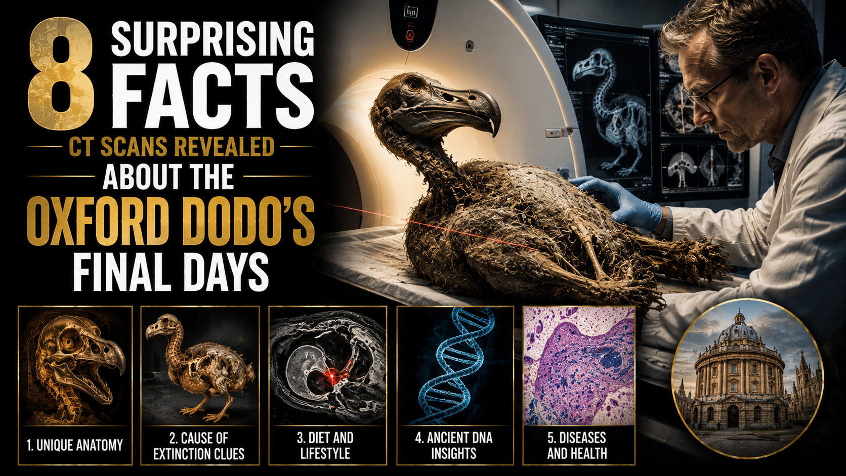
The dodo has long been one of the most famous symbols of extinction. Once native to Mauritius, this flightless bird disappeared just a few centuries after humans first encountered it. Today, much of what we know about the dodo comes not from living observation, but from fossils, historical accounts, and increasingly, modern imaging technology.
One of the most significant specimens is the Oxford dodo, preserved at the University of Oxford’s Natural History Museum. Unlike most ancient remains studied only through surface observation, this specimen has been examined using advanced CT scanning techniques, allowing scientists to look inside its bones without damaging them.
These scans have revealed surprising details about the bird’s biology, health, and final moments in a rapidly changing environment.
Here are eight surprising facts CT scans revealed about the Oxford dodo’s final days.
1. The Dodo’s Bone Structure Shows It Was a Powerful Bird
CT imaging confirmed that the dodo had a strong skeletal structure, particularly in its legs and pelvis.
These features suggest it was well adapted for walking and stability rather than flight.
Its bones were dense and robust, supporting a large body weight.
This reinforces the idea that the dodo was a ground-dwelling species built for strength rather than speed.
2. Signs of Stress Appeared in Its Growth Patterns
CT scans revealed irregularities in bone growth.
These patterns suggest the bird may have experienced periods of stress, possibly due to environmental changes or limited food availability.
Growth disruptions can indicate poor nutrition or habitat instability.
This provides clues about the challenges the species faced before extinction.
3. The Dodo’s Skull Structure Shows Specialized Feeding Habits
Detailed imaging of the skull revealed adaptations for a specific diet.
The shape of the beak and jaw muscles suggests the dodo was capable of processing hard fruits and seeds.
This specialized feeding behavior may have made it vulnerable when its environment changed.
Limited dietary flexibility often contributes to species decline.
4. CT Scans Reveal Muscle Attachment Points
Even without soft tissue, CT scans can identify where muscles once attached to bones.
The Oxford dodo shows strong attachment areas in the neck and legs.
This indicates it had powerful movements for walking and foraging.
These structural features help reconstruct how the bird moved in life.
5. The Bird Likely Lived in a Stable but Isolated Ecosystem
Bone analysis suggests the dodo evolved in a relatively stable island environment.
Isolated ecosystems often lead to unique adaptations due to lack of predators.
However, such isolation can also make species vulnerable to sudden environmental changes.
This helps explain why the dodo struggled after human arrival.
6. There Are No Clear Signs of Severe Injury
CT scans did not reveal major traumatic injuries in the Oxford specimen.
This suggests the bird likely did not die from a violent encounter.
Instead, its death may have been linked to environmental pressures or ecological disruption.
This challenges earlier assumptions that focused on direct predation alone.
7. The Skeleton Shows Adaptation for a Ground-Based Lifestyle
The structure of the legs and hips confirms the dodo was entirely flightless.
Its body was designed for walking long distances and navigating forest floors.
The absence of strong flight muscles aligns with this adaptation.
This evolutionary trade-off worked well in predator-free environments but became a disadvantage later.
8. The Scans Help Reconstruct the Dodo’s Final Ecological Challenges
Perhaps the most important insight is what the scans suggest about the bird’s environment.
Changes in habitat, introduced species, and human activity likely contributed to its decline.
While CT scans cannot show behavior directly, they reveal biological stress markers consistent with environmental pressure.
This helps build a more complete picture of the dodo’s final centuries.
Why the Oxford Dodo Still Matters Today
The Oxford dodo is more than a museum specimen—it is a scientific archive of a lost species.
Through modern CT scanning, researchers can study internal structures that would otherwise remain hidden.
This allows scientists to reconstruct not just what the dodo looked like, but how it lived and struggled in its environment.
It also serves as a powerful reminder of how quickly species can disappear when ecosystems are disrupted.
The Role of Technology in Understanding Extinction
CT scanning has revolutionized the study of extinct species.
Unlike traditional methods, it allows non-invasive analysis of fragile fossils.
This preserves the specimen while revealing internal details such as bone density, growth patterns, and structural adaptations.
As technology improves, even more insights may emerge from the Oxford dodo and similar specimens.
Final Thoughts
The CT scan analysis of the Oxford dodo provides a rare and detailed look into the life and final days of one of history’s most iconic extinct birds.
From its strong skeletal structure to subtle signs of environmental stress, each discovery adds depth to our understanding of its existence.
While the dodo itself is long gone, modern science continues to bring its story back to life.
In the end, the Oxford dodo reminds us that extinction is not just a historical event—it is a continuing lesson about the balance between species and their environment.